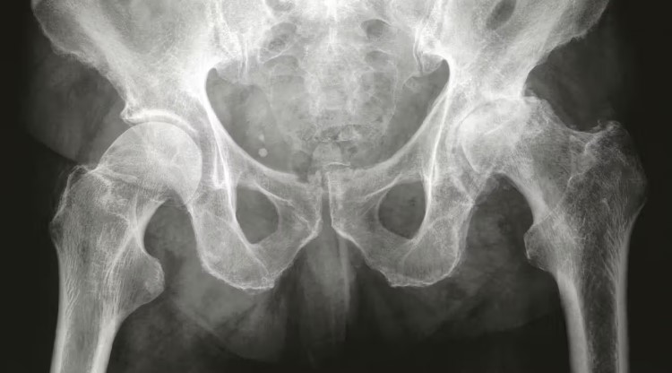
X-rays are a valuable tool that doctors may use to diagnose osteoarthritis. However, they may not always detect the condition, so physicians often consider additional information. Osteoarthritis occurs when cartilage in a joint wears down, leading to the contact of bones within the joint. This can result in pain and stiffness. Recent research suggests that the causes of osteoarthritis are more complex than initially thought.
In the hip, osteoarthritis typically manifests where the femur meets the pelvis. Normally, cartilage protects this area, but with osteoarthritis, the cartilage can deteriorate, resulting in pain and damage to the hip joint.
Continue reading to learn more about the appearance of osteoarthritis on an X-ray, what to expect during an X-ray procedure, and other diagnostic tests that doctors may use for diagnosing hip osteoarthritis.
What Does Osteoarthritis Look Like on an X-ray?
Your hip joint is a ball-and-socket joint. The ball-shaped, cartilage-covered femoral head fits into a socket in the pelvic bone, allowing your leg to move in various directions.
In a typical hip X-ray, you’ll see a space between the femoral head and the pelvis, which is where the cartilage cushions the femur in the joint.
If you have hip osteoarthritis, this joint space may appear significantly narrower because the cartilage has worn away, bringing the femoral head closer to the pelvic bone.
This causes the bones to scrape against each other as you move your leg, leading to significant pain and stiffness when walking, standing, or sitting. You may also observe cracks in the bone, missing pieces from the femoral head, or areas where the femur has hardened (subchondral sclerosis).
Damaged cartilage and bone fragments resulting from wear and tear may also be visible on an X-ray as white patches around the joint. Additionally, you may notice bone spurs or cysts that have developed on your bones due to joint disease.
An X-ray may also reveal chondrocalcinosis, which is the buildup of calcium crystals in the joint. This common complication of osteoarthritis appears as white lines on the surface of the cartilage.
Can an X-ray Detect the Severity of Osteoarthritis?
An X-ray can show wear and damage in the hip resulting from osteoarthritis.
A hip X-ray is often able to demonstrate:
- The extent of cartilage wear
- The narrowing of the joint space
- The damage to the femoral head
However, it is important to note that an X-ray may not always show cartilage damage from osteoarthritis.
What Is the Procedure for a Hip X-ray?
Hip X-rays are typically performed in radiology labs or clinics specializing in imaging tests.
You don’t need extensive preparation for a hip X-ray. Here are some tips to help make the X-ray process more comfortable:
- Wear loose clothing that is comfortable and easy to take on and off, especially if you need to change into a gown.
- Remove any jewelry or metal accessories as they may interfere with the X-ray images.
- Inform the technician if you have any metal implants in your body, as these may affect the X-ray results.
Some X-rays require you to stand next to a plate-shaped tool that captures the X-ray images. Other times, you may need to lie down so the technician can maneuver a specialized camera over your hip to take the images.
You must stand or lie very still while the X-ray image is taken to ensure clarity. Once the technician acquires the necessary images, you can change back into your regular clothes and leave the facility shortly afterward.
You may be able to review the X-ray results with a radiologist or doctor immediately. However, you might also need to schedule a follow-up appointment to discuss the results and potential treatments.
How Reliable Are X-rays for Detecting Hip Osteoarthritis?
According to research from 2022, X-ray images can’t always confirm a diagnosis of hip osteoarthritis, especially in the early stages.
On an X-ray, doctors may not always detect early structural changes, such as joint space narrowing and damage to the femoral head. These early changes are often when you first start experiencing noticeable pain and alterations in your gait. Many people experiencing this pain don’t show visible signs on an X-ray.
Therefore, doctors need to use X-rays in conjunction with other diagnostic tests to assess the progression of osteoarthritis and the extent of joint damage. This comprehensive approach helps them choose an appropriate treatment option, such as hip arthroplasty.
What Other Tests Do Doctors Use to Detect Hip Osteoarthritis?
In addition to X-rays, doctors may employ several other tests and procedures to detect hip osteoarthritis, including:
- Physical exam: Assessing the joint’s range of motion and tenderness.
- Symptom discussion: Discussing your pain levels and mobility issues.
- Review of risk factors: Evaluating your age, weight, and family history.
- Comparison of leg lengths: Identifying any discrepancies that may indicate joint issues.
- Gait analysis: Observing how you walk to detect any abnormalities.
- Additional imaging: Utilizing magnetic resonance imaging (MRI) or computed tomography (CT) scans to examine the nerves and blood vessels around the joint.
- Blood tests: Checking for signs of rheumatoid arthritis (RA).
Takeaway
Doctors typically perform hip X-rays and other diagnostic tests to identify signs of wear and tear in the hip.
Osteoarthritis is relatively easy to manage if detected early. If you are experiencing significant pain and stiffness while walking, contact a doctor to get X-ray imaging and other tests to determine the appropriate course of treatment.
